Antenatal Hydronephrosis
What is antenatal hydronephrosis?
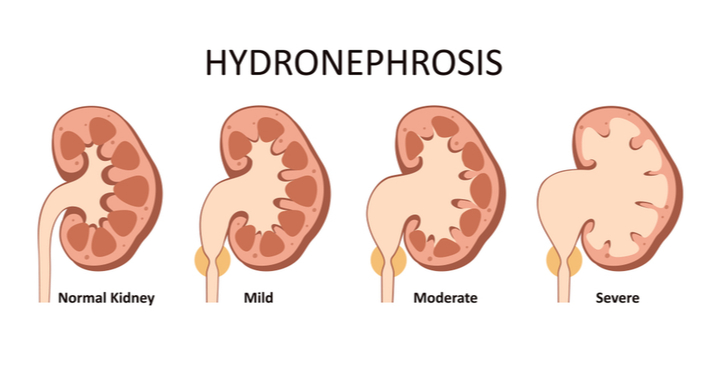
Antenatal hydronephrosis occurs when the kidney enlarges and fills with fluid before birth. Ultrasound examinations may identify it in the fetus as early as the first trimester of pregnancy. Most of the time, this diagnosis will not affect prenatal treatment, but it may need monitoring and perhaps surgery throughout infancy and childhood.

What factors contribute to prenatal hydronephrosis?
The following are some of the possible causes of prenatal hydronephrosis:
- Blockage may occur at the Ureteropelvic junction (UPJ/ PUJ) of the kidney, at the bladder at the ureterovesical junction, or in the urethra (posterior urethral valves).
- Reflux occurs when the valve between the bladder and the ureter malfunctions, allowing urine to flow back up to the kidney as the bladder fills or empties. Most children (75%) overcome this throughout childhood, however they need daily antibiotic prophylaxis to avoid kidney damage before they outgrow the reflux.
- Duplications: around 1% of all persons have two collecting tubes from their kidney. These may be seen on prenatal ultrasonography. Patients with duplication may develop a ureterocele, which is a balloon-like blockage at one of the duplex tubes.
- Multicystic kidney: This is a nonfunctional cystic kidney.
- Many of these dilated kidneys return to normal after birth, which is known as transient hydronephrosis.
- It is advisable to visit a pediatric urologist as soon as the child is diagnosed with fetal hydronephrosis in order to plan for effective treatment after delivery.
What is done to evaluate the hydronephrosis after the baby is born?
To examine the kidneys, many investigations may be required:
- During the newborn period, ultrasound is used to assess the size and edema of the kidneys, ureter, and bladder.
- A voiding cystourethrogram is done in cases where PUV or vesicoureteral reflux is suspected.
- Diuretic renal scan to evaluate kidney function and drainage, particularly in cases of PUJ obstruction; DMSA scan to check the cortical function of the kidney in selected cases of VUR; CT-IVP- in case of an anatomical issue to delineate clear anatomy; but is rarely required these days due to good USG and renal scans.
How is antenatal hydronephrosis managed during pregnancy?
Ultrasound testing is all that is required in virtually all cases of prenatal hydronephrosis.
Draining the kidneys or bladder by tube or surgery has been done experimentally in the uncommon baby with significant obstruction of both kidneys and inadequate amniotic fluid (the fluid surrounding the fetus). While these procedures are technically possible, the outcome of these babies has not been improved. These babies are likely to have severely dysfunctional kidneys that do not function properly, as well as inadequate lung development. Most instances of prenatal hydronephrosis have no effect on pregnancy and may be delivered normally. Large blocked kidneys may need a C-section, although this is uncommon.
Is fetal hydronephrosis serious?
Most of the time, fetal hydronephrosis is not serious, but pregnant women must have regular ultrasounds to see whether the problem has improved or if it needs care after birth.
Treatment for Antenatal Hydronephrosis
Prior to birth– In most circumstances, no therapy is required prior to birth. A procedure during pregnancy may be appropriate in a very limited percentage of circumstances.
Following birth– This is determined by the results of prenatal ultrasound scans and postnatal examinations. Most newborns may be discharged home within a few hours after delivery.
Rarely, newborns are born with difficulties and must be sent to a neonatal unit, a section of the hospital dedicated to newborn babies, for monitoring and care.
Preventing and treating UTIs-Preventing and treating UTIs If they keep recurring, they may cause kidney damage. To preserve the kidneys, it is critical to avoid UTIs and treat them as soon as they occur.
Other therapies– Only a small percentage of newborns need further therapy, like as surgery, to repair the condition that caused the hydronephrosis. A pediatric urologist will explain what will happen if your infant requires treatment.
For more information & consultation on Antenatal Hydronephrosis, Get in touch with Dr. Adwait Prakash a Pediatric Surgeon in Indore. will help you out in understanding your problem and guide you through every stage of your treatment.
To book your appointment Call: 8889588832.
