Anorectal Malformation
Anorectal Malformation
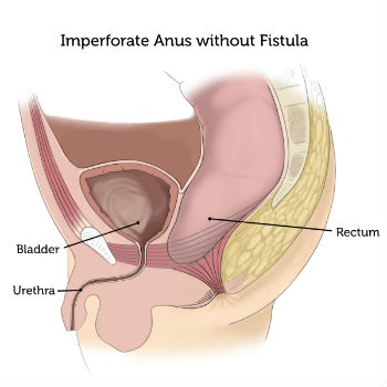
Anorectal malformations are birth defects (problems that happen as a fetus is developing during pregnancy). With this defect, the anus and rectum (the lower end of the digestive tract) do not develop properly.

What are the different types of Pediatric Anorectal Malformation (Imperforate Anus)?
- Anorectal anomalies are classified into numerous forms, including:
- Narrow anal passages
- The rectum does not connect to the anus
- Anus imperforate
- Cloacal abnormality
What are the indications and symptoms of an imperforate anorectal malformation in children?
Anorectal abnormalities in neonates make it difficult for a baby to produce a bowel movement. Bloating, difficulty or inability to make a bowel movement, and fever may occur in children.
Who is at risk for developing the disorder?
The etiology of an anorectal malformation is usually unknown.
- Some of the following genetic disorders or congenital issues may cause anorectal malformation:
- The VACTERL association (a syndrome in which there are Vertebral, Anal, Cardiac, Tracheal, Esophageal, Renal, and Limb abnormalities)
- Abnormalities in the digestive system
- Abnormalities of the urinary tract
- Abnormalities of the spine
What is the diagnostic test for anorectal malformations?
When your baby is delivered, the doctor will do a physical examination and check to see whether the anus is open. To further examine the issue, diagnostic imaging studies such as:
- Abdominal X-rays. A diagnostic technique that employs invisible electromagnetic energy beams to create images on film of inside tissues, bones, and organs.
- Abdominal ultrasound (also called sonography). A diagnostic imaging technology that creates pictures of blood arteries, tissues, and organs using high-frequency sound waves and a computer. Ultrasounds are used to see into organs and to measure blood flow via different arteries.
- Computed tomography scan -A diagnostic imaging process that use X-rays and computer technology to generate horizontal, or axial, images of the body. A CT scan provides comprehensive pictures of any region of the body, such as the bones, muscles, fat, and organs. CT scans provide more information than standard X-rays.
- Magnetic resonance imaging (MRI). A diagnostic process that produces comprehensive images of organs and structures inside the body using a combination of massive magnets, radio frequencies, and a computer.
- Lower gastrointestinal (GI) series (also called barium enema). The rectum, large intestine, and lower section of the small intestine are all examined during a lower GI series. A fluid called barium (a metallic, chalky, liquid used to coat the inside of organs so that they will show up on an X-ray) is given into the rectum as an enema. An abdominal X-ray reveals strictures (narrowed regions), obstructions (blockages), and other issues.
- Upper gastrointestinal (GI) series (also called barium swallow). Upper GI series is a diagnostic test that examines the organs of the upper part of the digestive system: the esophagus, stomach, and duodenum (the first section of the small intestine). Barium fluid (a metallic, chalky substance used to coat the interior of organs so they show up on an X-ray) is swallowed. The digestive organs are next evaluated using X-rays.
What are the treatment options for Anorectal Malformation?
A colostomy is a surgical procedure that provides an incision for the colon, or large intestine, to excrete waste into a small bag outside of the body. Within the first few days of life, most newborns born with an anorectal abnormality will need a temporary colostomy. The colostomy ensures that the newborn can pass stool normally, helps to regulate digestion, and greatly minimizes the risk of infection.
Anorectal reconstructive surgery, also known as posterior sagittal anorectoplasty (PSARP), is done to connect the rectum to the anal hole and cover any aberrant openings that may interfere with the ability to have a regular bowel movement. In cases where there is an absence of an anal opening, your surgeon will create a new one.
The majority of PSARP treatments are done on babies aged one to six months. The kind of PSARP done will be determined by the location of the rectum and anus (low or high), the function of the sphincter muscles, and the presence of any aberrant holes (called fistulas) that need to be fixed.
Colostomy Closure
Your child’s colostomy will remain in place for approximately eight weeks following the reconstructive surgery to allow the rectum and anal opening to heal before coming in contact with any waste. The colostomy will be medically closed when your child is ready and totally recovered. Within a few days, they will be able to pass stools on their own via the anus. Initially, stools will be loose and frequent. Safe topical remedies should be used to protect your child’s skin against diaper rash and discomfort. Your child’s stools will normalize as they recover, becoming firmer and less frequent.
For more information & consultation on Anorectal Malformation, Get in touch with Dr. Adwait Prakash a Pediatric Surgeon in Indore. will help you out in understanding your problem and guide you through every stage of your treatment.
To book your appointment Call: 8889588832.
