Spina Bifida
What is Spina Bifida
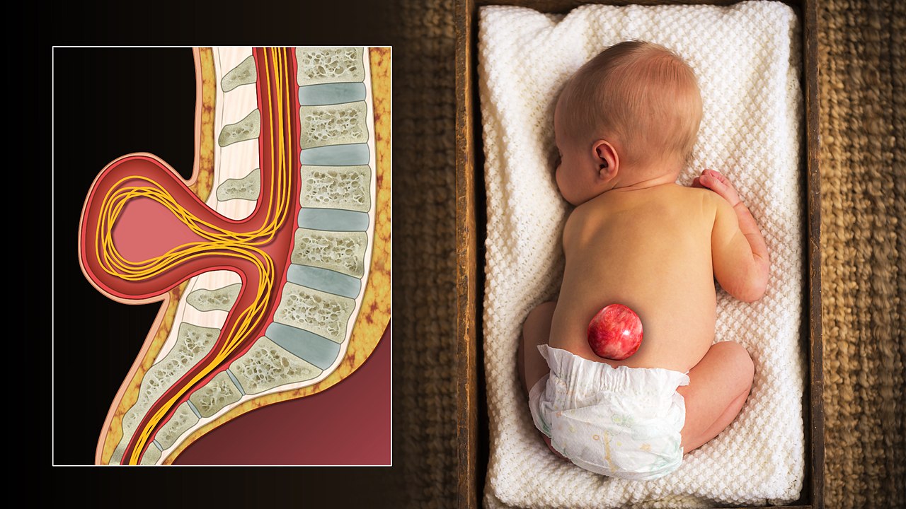
Spina bifida is a condition in which a baby’s spine and spinal cord do not grow correctly in the womb, resulting in a gap in the spine. A neural tube defect is spina bifida. The neural tube is the structure from which the baby’s brain and spinal cord grow. The neural tube develops early in pregnancy and closes approximately 4 weeks after conception.
Spina bifida occurs when a portion of the neural tube fails to form or shut correctly, resulting in abnormalities in the spinal cord and spine bones (vertebrae).

Spina Bifida Causes
Although the etiology of spina bifida is unknown, a number of variables might raise a baby’s risk of developing the condition.
These include:
- Folic acid deficiency during pregnancy
- Genetic causes
- Taking certain medications during pregnancy, such as valproic acid (used to control seizures), has been associated with an increased chance of having a baby with spina bifida.
The symptoms of spina bifida depend on exactly where and what extent the spinal cord and overlying structures have not developed correctly. There are three common subtypes:
Occulta is often called hidden spina bifida, as the spinal cord and the nerves are usually normal and there is no opening on the back. This kind of spina bifida has just a minor defect or gap in the small bones (vertebrae) that make up the spine, and it affects roughly 12% of the population. In many situations, spina bifida occulta is so mild that no spinal function is affected. Unless it is identified on an x-ray, most individuals are unaware that they have spina bifida occulta. However, one in every 1,000 people has an occult structural discovery that causes neurological impairments or disabilities such as bowel or bladder problems, back discomfort, leg weakness, or scoliosis.
Meningocele arises when the bones surrounding the spinal cord do not close properly and the meninges are forced out through the opening, resulting in the formation of a fluid-filled sac. The dura mater, arachnoid mater, and pia mater are three layers of membranes that envelop the spinal cord. In most instances, the spinal cord and nerves are unaffected or very mildly impacted. The sac is often covered by skin and may need surgery. This is the most uncommon kind of spina bifida.
Myelomeningocele accounts for about 75% of all spina bifida cases. The most severe version of the condition occurs when a part of the spinal cord itself protrudes through the back. In some situations, skin covers the sacs, while in others, tissue and nerves are exposed. The degree and location of the spinal cord abnormalities determine the level of neurological impairments. If just the bottom of the spinal cord is affected, there may be only bowel and bladder problems, but more severe instances might result in complete limb paralysis with bowel and bladder distress.
Testing & Diagnosis
- Infant ultrasound
- MRI of the fetus
- Alpha-fetoprotein level in maternal serum ( tests for confirmation including amniocentesis)
- Ultrasound at 20 weeks
Treatment
Prenatal
Pregnant women of reproductive age may minimize their chance of having a child with spina bifida by taking 400 micrograms (mcg) of folic acid daily. Folic acid, since it is water soluble, does not remain in the body for long and must be taken on a daily basis to be effective against neural abnormalities. Because half of all pregnancies are unplanned, folic acid should be taken whether or not a woman intends to get pregnant. According to research, if all women of reproductive age took a multivitamin containing the B-vitamin folic acid, the incidence of neural tube abnormalities might be decreased by up to 70%.
Fetal Surgery
A randomized trial (MOMS Trial) compared to repair of the defect while the baby was still in the uterus to repair after delivery, and the outcomes were hopeful but variable. Patients who had fetal repair were less likely to need a ventriculo-peritoneal shunt (44 percent vs. 84 percent) and more likely to be able to walk (44 vs. 24 percent ). The fetal surgery group also had many more maternal and infant complications.
Infant Surgery
A baby born with spina bifida needs to have the exposed part of the spinal cord repaired to prevent further injury and infection. A neurosurgeon reintroduces the neural tissues into the spinal canal before closing the muscle and skin. This surgery performed a few hours after delivery, was formerly considered a medical emergency. Most surgeries are now finished within the first 48 hours of a baby’s life.
Hydrocephalus affects 80 to 90 percent of children with spina bifida. Hydrocephalus is a disorder in which excess cerebrospinal fluid (CSF) accumulates inside the ventricles (fluid-containing chambers) of the brain, potentially increasing head pressure. To manage the buildup of spinal fluid, the majority of these children will need a ventricular shunt. The shunt will stay in place for the rest of the person’s life, although it will need to be changed several times.
Dr. Adwait Prakash is an expert in Spina Bifida Treatment practicing in Indore.
To book your appointment Call: 8889588832
